Types of Dermatomyositis
Dermatomyositis is classified into different subtypes based on the extent of skin or muscle involvement:
Juvenile dermatomyositis occurs in patients < 18 years old at onset3
Juvenile dermatomyositis is the most common inflammatory myopathy of childhood. Juvenile Dermatomyositis is characterized by the presence of the typical rash and proximal muscle weakness. Compared to dermatomyositis in adults, pediatric dermatomyositis patients show greatest weakness in hip flexors, extensors, and adductors, as well as neck flexors and shoulder abductors. Characteristic dermatologic features of juvenile dermatomyositis involve the heliotrope rash, Gottron’s papules, and nail fold changes. Calcinosis is a common finding (up to 40% of patients) in juvenile dermatomyositis.6
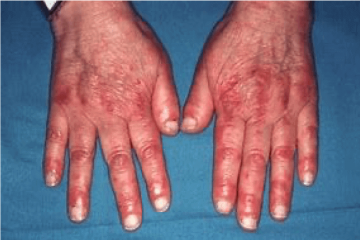
Image shows Gottron's signs on hands. Gottron’s signs are skin changes that may be seen in people living with dermatomyositis. These signs are typically observed on the knuckles and fingers and may include redness of the skin (Erythema).2
References
- Goyal, N. A. (2019). Immune-Mediated Myopathies. CONTINUUM: Lifelong Learning in Neurology, 25(6), 1564–1585. https://doi.org/10.1212/con.0000000000000789
- Dalakas, M. C., & Hohlfeld, R. (2003). Polymyositis and dermatomyositis. The Lancet, 362(9388), 971–982. https://doi.org/10.1016/s0140-6736(03)14368-1
- Malik, A., Hayat, G., Kalia, J. S., & Guzman, M. A. (2016). Idiopathic Inflammatory Myopathies: Clinical Approach and Management. Frontiers in Neurology, 7, 64. https://doi.org/10.3389/fneur.2016.00064
- Callander, J., Robson, Y., Ingram, J., & Piguet, V. (2016). Treatment of clinically amyopathic dermatomyositis in adults: a systematic review. British Journal of Dermatology, 179(6), 1248–1255. https://doi.org/10.1111/bjd.14726
- Schmidt, J. (2018). Current Classification and Management of Inflammatory Myopathies. Journal of Neuromuscular Diseases, 5(2), 109–129. https://doi.org/10.3233/jnd-180308
- Swafford, C., and Roach, E.S. (2020). Juvenile Dermatomyositis and the Inflammatory Myopathies. Semin. Neurol. 40, 342-348. https://doi.org/10.1055/s-0040-1705120
Science Hub
Sign up and connect to Science Hub to learn more about our recent ProDERM study and have access to symposia presentations, publications, posters and lectures.